When a dog limps or hesitates before jumping onto the couch, most owners immediately reach for supplements without understanding what's actually occurring inside the joint. That gap in knowledge leads to poor product choices, missed warning signs, and delayed intervention. Understanding the basic anatomy of canine joints is the foundation of every effective joint health strategy I've developed over 12 years.
This isn't a veterinary textbook. It's a practical explanation of how joints work, why they degrade, and what that means for the supplements and management strategies that actually help.
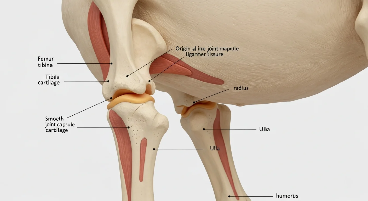
The Components of a Canine Synovial Joint
Most of the joints that concern dog owners — hips, elbows, knees, shoulders — are synovial joints. They share the same fundamental architecture regardless of location.
Articular Cartilage: The Shock Absorber
Cartilage is the smooth, white tissue that covers the ends of bones where they meet. It has two jobs: reducing friction between bone surfaces and absorbing compressive forces during weight-bearing activity. A healthy dog's articular cartilage is roughly 70% water, held in a matrix of collagen fibers and proteoglycans. This matrix gives cartilage its remarkable ability to deform under load and spring back when pressure is released.
The critical detail most supplement marketing ignores: cartilage has no blood supply. It receives nutrients by diffusion from the synovial fluid surrounding it. This is why injury and inadequate joint fluid quality so severely compromise cartilage health — and why anti-inflammatory nutrition that supports synovial fluid quality matters so much.
Synovial Fluid: The Joint's Lubrication System
Synovial fluid is produced by the synovial membrane that lines the joint capsule. It serves two functions: lubricating the cartilage surfaces to reduce friction and delivering nutrients to the avascular cartilage. Healthy synovial fluid is thick and viscous, like egg white. Inflamed joints produce thinner, less effective fluid that fails to adequately lubricate or nourish the cartilage.
Hyaluronic acid is the primary molecule responsible for synovial fluid viscosity. This is why injectable hyaluronic acid can be effective for joint disease — the molecule is delivered directly where it's needed. Oral hyaluronic acid supplements are far less convincing because the large molecules are unlikely to survive digestion and reach the joint intact.
Subchondral Bone: The Foundation
Beneath the cartilage sits the subchondral bone, which acts as the structural foundation. In early osteoarthritis, this bone begins to thicken and harden in response to increased stress from degrading cartilage above it. The subchondral bone contains nerve endings, which is why severe arthritis — where cartilage has largely worn away — causes bone-on-bone pain.
The Joint Capsule and Ligaments
The entire joint is enclosed in the joint capsule, a fibrous structure that maintains joint integrity and contains the synovial membrane. Ligaments connect bone to bone and provide passive stability. Tendons connect muscles to bones and transmit the forces that move the joint. Injuries to these soft tissues often contribute to joint instability that accelerates cartilage wear.
How Osteoarthritis Develops: The Cascade
Osteoarthritis (OA) isn't simply "wear and tear." It's an active disease process involving cartilage breakdown, bone remodeling, and chronic low-grade inflammation. Understanding the sequence helps you understand why early intervention matters.
Stage 1: Cartilage Matrix Disruption
The first change is often subtle — the proteoglycan matrix that holds water in cartilage becomes less organized. Water content rises, and the cartilage becomes softer and more vulnerable to mechanical damage. This stage is usually invisible on standard X-rays. The dog may show no obvious symptoms, which is why recognizing subtle early signs requires attention to behavioral changes rather than waiting for visible lameness.
Stage 2: Synovial Inflammation
As cartilage breaks down, degradation products enter the synovial fluid. The synovial membrane responds by producing inflammatory mediators — cytokines and proteases that paradoxically accelerate further cartilage destruction. This creates a self-perpetuating inflammatory cycle. Omega-3 fatty acids, particularly EPA, work at this stage by competing with arachidonic acid in the inflammatory pathway. This is why consistent omega-3 supplementation is one of the most evidence-supported interventions available.
Stage 3: Structural Remodeling
In established OA, the body attempts to stabilize the joint through bony remodeling. Osteophytes — bone spurs — form at joint margins. Subchondral bone thickens. The joint capsule may thicken and fibrosis. These changes restrict range of motion and contribute to the morning stiffness characteristic of arthritic dogs. At this stage, physical management becomes as important as supplementation. The exercise modification guide covers how to adapt activity levels to match each stage of joint disease.
Why Large Breed Dogs Are More Vulnerable
Body weight creates compressive forces on joints that scale with mass. A 100-pound dog doesn't just have twice the joint stress of a 50-pound dog — because of the physics of force per unit area, the relationship is less linear than that, but the principle holds. Large breed dogs also tend to have faster growth rates that can outpace the development of supporting musculature and connective tissue.
Hip dysplasia and elbow dysplasia, the most common orthopedic conditions in large breeds, involve malformation of the joint socket or incongruity between joint surfaces. These create abnormal contact patterns that accelerate cartilage wear in specific locations. Breeds like German Shepherds, Labrador Retrievers, Golden Retrievers, and Rottweilers carry significantly elevated genetic risk. For these dogs, proactive supplementation should begin well before clinical signs appear, as detailed in the age-by-age joint protocol guide.
The Role of Muscle in Joint Health
This is chronically underemphasized in supplement discussions. Muscles are the primary active stabilizers of joints. Strong, well-conditioned muscles reduce the load that joint capsules, ligaments, and cartilage must bear. A dog with good muscle mass distributes weight-bearing forces more effectively and protects joints from impact and shear forces.
Atrophy of muscles surrounding a diseased joint is a vicious cycle — the dog avoids using the painful limb, the muscles weaken, the joint loses its primary stabilizer, and the disease progresses faster. Appropriate exercise and physical therapy aren't just comfortable additions to a joint health program; they're mechanically essential. This is why I push back against owners who believe they can address joint disease with supplements alone without managing activity levels.
Connective Tissue Turnover: Why Timing Matters
Cartilage matrix components have turnover rates measured in weeks to months. Collagen fibers in cartilage are particularly slow to replace. This is why joint supplement trials need to run for a minimum of 8 to 12 weeks before you can assess whether they're working. The rapid improvements sometimes reported within days of starting a supplement are almost certainly placebo effect or natural disease fluctuation, not cartilage repair.
Understanding this turnover also clarifies when to start supplementation. If you begin supporting cartilage matrix synthesis in a young dog before significant degradation has occurred, the maintenance requirements are much lower. Once significant cartilage is lost, no supplement can restore it — you're managing what remains, not rebuilding what's gone. The joint supplements science guide covers which ingredients address which components of this biology.
Practical Implications for Your Supplement Choices
With this anatomy in mind, here's how to think about the major categories:
- Glucosamine and chondroitin provide substrates for cartilage matrix synthesis and may reduce enzymatic breakdown of proteoglycans.
- Omega-3 fatty acids (EPA/DHA) reduce synovial membrane inflammation, improving fluid quality and breaking the inflammatory cycle.
- Undenatured type II collagen (UC-II) works via immune modulation to reduce antibody-mediated cartilage destruction — a different mechanism from the structural supplements above.
- Green-lipped mussel combines glycosaminoglycans with unique omega-3s, addressing both structural and inflammatory components.
No supplement reverses established structural damage. The goal is slowing progression, managing inflammation, and supporting the remaining cartilage. Combined with appropriate weight management, exercise, and diet, that's achievable for the majority of arthritic dogs.