
If your vet has ordered imaging for your dog's joints or bones, the short answer is this: an X-ray image is a shadow cast by tissue onto a detector, a CT scanner builds a slice by computing X-ray measurements taken from many angles, and an MRI scan uses magnetic fields and radio waves instead of X-rays, so it involves no ionising radiation at all. The three methods answer different questions, and your vet picks between them based on what needs to be seen.
What is an X-ray image, and why is it like a shadow?
An X-ray tube fires a beam of high-energy photons through the dog's leg, hip, or spine. Dense tissue, mainly bone, absorbs more of that beam. Soft tissue absorbs less. Air absorbs almost none. Whatever passes through lands on a detector on the far side and forms a two-dimensional image.
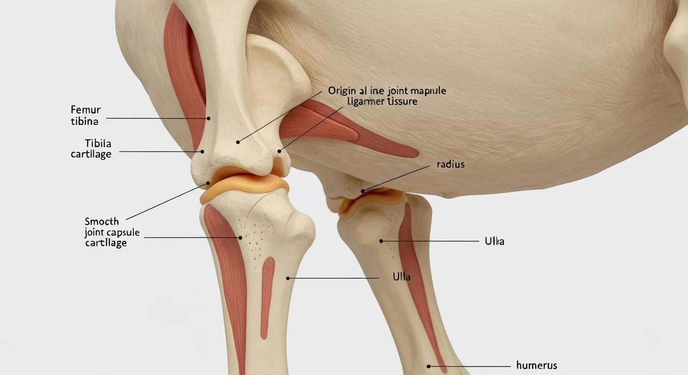
The shadow analogy is not a metaphor, it is the actual mechanism. Put your hand between a lamp and a wall and you get a flat, overlapping silhouette. An X-ray image works the same way: everything in the beam's path is superimposed on one plane. A hip joint, the femur head, the acetabulum, and any soft tissue in front of or behind them all land on the same picture. That is why radiographers take at least two views, usually a side view and a front-to-back view, so the vet can mentally separate structures that overlap on a single image.
Bone shows up pale because it blocks the beam. Gas in the stomach or bowel shows up dark. Mineralised joint bodies, bone fragments, and osteoarthritis changes such as new bone at the joint margins are usually visible. Early cartilage loss is not, because cartilage is soft tissue and does not cast a distinct shadow. This is the main limitation of radiography for joints, and it is why your vet may move to a different method. If you want to understand the same principles in human medicine, from the written request to the final report, this plain-English explainer of X-ray image, shadow and the other imaging methods covers the whole pathway.
For a dog, positioning matters as much as the machine. Most general practices take survey radiographs with the dog awake or lightly sedated. Orthopaedic views, such as those used for hip scoring, often need general anaesthesia so the limbs can be held in a precise, repeatable position. A slightly rotated pelvis can change how a joint looks, so the person holding or positioning the dog is doing real technical work.
How does a CT scanner create a computed slice?
A CT scanner is an X-ray tube and a detector ring that rotate around the dog while the table moves through the gantry. Instead of one flat shadow, the machine collects hundreds of transmission measurements from many angles around the same body region.
A computer then solves a reconstruction problem: given all those overlapping measurements, what distribution of tissue density would produce them? The output is a stack of thin cross-sectional images, each one a slice through the body. "Computed" in computed tomography refers to exactly this step. The slice is not photographed, it is calculated.
For canine joints, CT is useful where overlapping bone hides the answer on a plain film. Examples include complex fractures, elbow dysplasia, fragmented coronoid process, and bone tumours where the vet needs to see the extent of the lesion and whether the cortex is breached. Because each slice is thin, usually under a millimetre in modern scanners, small fragments that would blur together on a radiograph can be separated.
CT uses ionising radiation, and doses are higher than for a single radiograph because the scanner is taking many projections rather than one or two. In veterinary practice this is managed the same way it is in human medicine: justify the examination, meaning confirm it will change the plan, and optimise it, meaning use the lowest dose that still answers the question. Dogs are usually anaesthetised for CT so they stay perfectly still, which also prevents repeat scans.
Why does an MRI scan not use ionising radiation?
MRI stands for magnetic resonance imaging, and it uses no X-rays. The scanner creates a strong static magnetic field. Hydrogen nuclei, mostly in water and fat, align with that field. A pulse of radio waves knocks them out of alignment, and as they relax back, they emit a faint signal that receiver coils pick up. The computer turns those signals into images.
Because the contrast comes from how much water and fat sit in different tissues, MRI is excellent for soft tissue: ligaments, tendons, cartilage, menisci, spinal cord, and the fluid inside a joint. A cranial cruciate ligament tear, a meniscal injury, or a disc extrusion in the spine can be seen directly, which is not possible on a plain radiograph. For a dog with a swollen joint and normal X-rays, MRI is often the next step.
The trade-off is practical rather than biological. MRI takes longer, sometimes 30 to 60 minutes per region, and the dog must be completely motionless, so general anaesthesia is standard. The magnet is loud, and metal implants, some microchips, and certain foreign bodies need to be screened beforehand. Cost is usually higher than CT. The absence of ionising radiation is a genuine advantage, particularly for repeated scans, but it is not the only reason a vet chooses it.
Which method will my vet choose for my dog?
Radiography is first for most orthopaedic complaints. It is fast, widely available, and good at showing bone, joint alignment, and mineralised changes. It is the standard starting point for hip dysplasia screening, osteoarthritis monitoring, and suspected fractures.
CT is chosen when bone detail matters and structures overlap: complex elbow or carpal injuries, surgical planning, tumour staging, and some spinal cases. It gives a three-dimensional dataset the surgeon can rotate and measure.
MRI is chosen when the problem is soft tissue: suspected cruciate or meniscal injury, spinal cord compression, inflammatory joint disease, or a joint that looks normal on X-ray but is clearly painful. Ultrasound is a fourth option, using sound echoes rather than X-rays, and is useful for tendons and fluid collections near the surface.
In practice, these are often sequential. A radiograph raises a question, CT or MRI answers it, and the report comes back to your vet with a description of what was seen and what it likely means.
What should I expect on the day?
Most imaging appointments follow a similar shape. Your vet writes a clinical request explaining the question to be answered, the practice reviews it, and the imaging team plans the study. If contrast medium is needed, it is given intravenously, and the dog is monitored for reactions. You will usually be asked to withhold food for several hours beforehand if anaesthesia is planned.
After the scan, a radiologist or the attending vet reviews the images and writes a report. Ask for the key findings in plain language: what was seen, how certain it is, and what it changes about treatment. If the report mentions a term you do not know, ask what it means for your dog's daily routine, because that is usually the part that matters most at home.
One practical note: keep copies of the images and reports. If you later see an orthopaedic specialist or a rehabilitation vet, having the original study saves repeating an anaesthetic and a dose of radiation. Imaging is a snapshot in time, and comparing an old study with a new one often tells the story better than either alone.
Background sources: NHS.